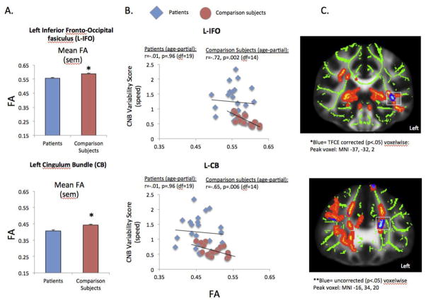
Figure 3.
Correlation between speed WIV and FA in comparison subjects and schizophrenia patients. A) Mean FA in patients and controls in the inferior fronto-occipital fasciculus (IFO) and cingulum bundle (CB); *p<.05. B) Correlation in the inferior fronto-occipital fasciculus (IFO) and cingulum bundle (CB) in patients and controls. FA values were extracted from the comparison>patient analysis from the JHU white matter tract regions-of-interest. C. Significant correlation between speed WIV and FA in healthy controls in a voxelwise analysis for the left inferior fronto-occipital fasciculus and left cingulum bundle (MNI coordinates reported for peak voxel). Regions are displayed with local enhancement for better viewing in radiological convention (right-left).

