
Figure 1.
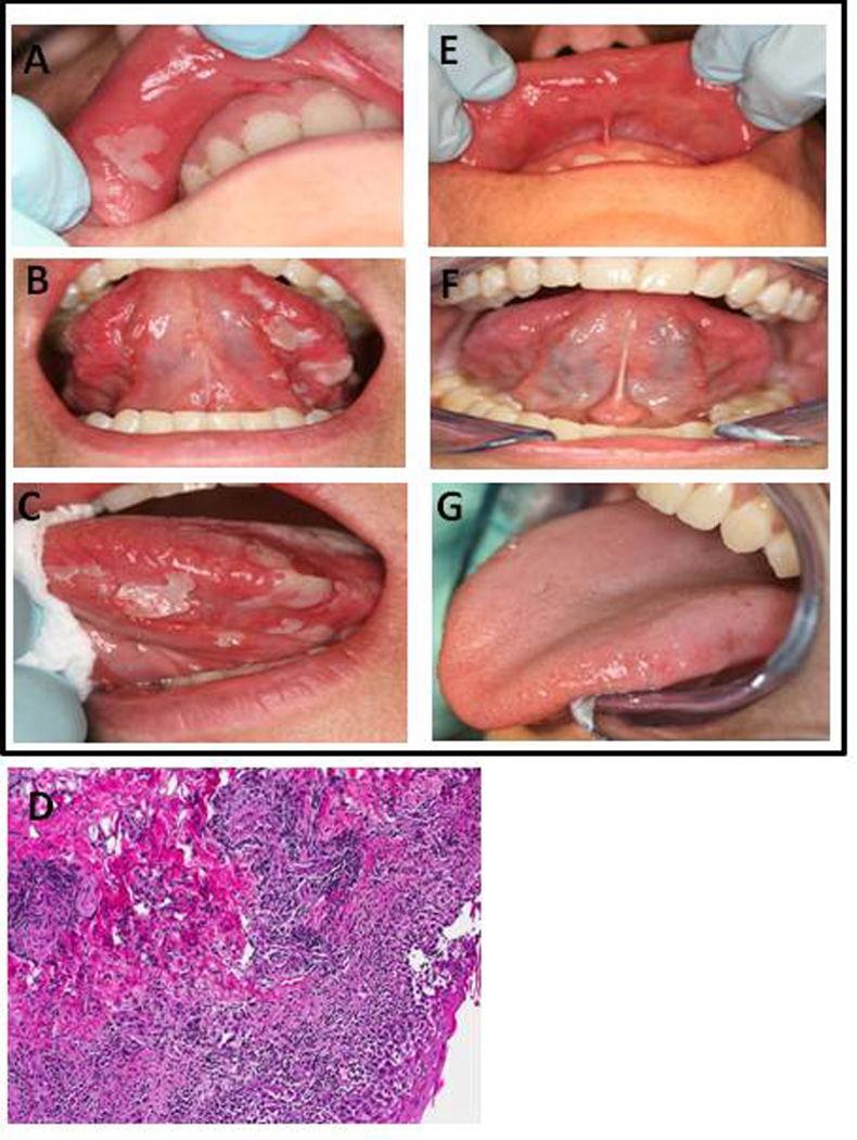
Clinical oral presentation includes (A, B, C) shallow ulcerations with an erythematous halo and pseudomembrane on the labial mucosa, and larger (1 cm) ulcerations on the tongue. (D) H&E staining of the upper labial mucosa reveals prevalent lymphocytic infiltrate and necrotic keratinocytes. Labial biopsy section shown at 10× magnification. (E,F,G) Results post-treatment with dexamethasone (0.5 mg/5 ml) oral rinse.