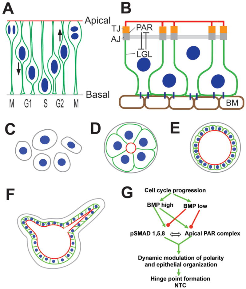
Figure 2. Epithelial organization and tissue morphogenesis.

(A) Cross-sectional/apicobasal view of the neural plate showing its pseudostratified epithelial organization and interkinetic nuclear migration. Cell nuclei are shown in blue, and the cell outline, including apical and basal processes, are shown in green. Based on Sauer et al., 1935; Baye and Link, 2007. (B) Neural epithelial cell organization with the apical (red) and basolateral (green) compartments separated by tight junctions (TJ). Adherens junctions (AJ) are located basal to the TJ and associate with the adherens/actin belt (grey). TJ and AJ are associated with the apical PAR complex, which antagonizes basolateral proteins, e.g., lethal giant larva (LGL). BM: basement membrane. (C–F) MDCK cells in 3D cultures (C) can self-organize to form cysts with central lumens (D). The cyst can expand by cell division (E), display dynamic variability in apicobasal polarity, leading to the formation of hollow tubes (F). C-F adapted from Mostov et al., 2003 and Zegers et al., 2003. (G) Cartoon depicting the role of BMP-apicobasal polarity interactions during hinge point formation. For details, see text. Adapted from Eom et al., 2012.
Abbreviations: AJ: adherens junctions; AO: area opaca; DLHP: dorsolateral hinge point; HN: Hensen’s node; LGL: lethal giant larva; MHP: median hinge point; NC: notochord; NF: neural fold; NP: neural plate; NT: neural tube; NTC: neural tube closure; PS: primitive streak; SE: surface ectoderm; TJ: tight junctions.
