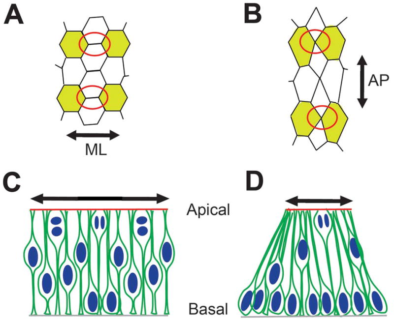
Figure 4. Convergent extension and cell cycle kinetics in neural tube closure.

(A, B) Neural plate cells in apical view showing progressive convergent extension. Apical junctional remodeling (red circles in A and B) results in mediolateral convergence and anteroposterior extension (double headed arrows in A and B, respectively). Note that such remodeling can produce apical constriction (Based on data from Nishimura et al., 2012). (C, D) Lateral neural plate cells display asynchronous cell cycle progression, with neighboring cells in different cell cycle phases (C). By contrast, a majority of nuclei at the MHP are basally located (D). Although the same number of cells is shown in C and D, the apical surface area in D is reduced compared to C (compare double headed arrows in C and D). Based on data from Schoenwolf et al., 1987; Eom et al., unpublished observations.
Abbreviations: AJ: adherens junctions; AO: area opaca; DLHP: dorsolateral hinge point; HN: Hensen’s node; LGL: lethal giant larva; MHP: median hinge point; NC: notochord; NF: neural fold; NP: neural plate; NT: neural tube; NTC: neural tube closure; PS: primitive streak; SE: surface ectoderm; TJ: tight junctions.
