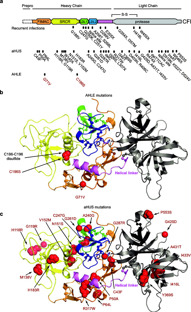
FIG. 3. Molecular modeling of CFI mutations.
a, Domain structure with the positions of mutations associated with recurrent infections, aHUS, and AHLE [4–6, 12–16]. Mutations are numbered inclusive of the signal peptide (+18 relative to position numbers when the signal peptide is excluded) [13]. b, Structure of human FI [17] with AHLE mutations (red spheres) c. Structure with aHUS mutations (red spheres). Molecular images generated by PyMol [41]. FIMAC, Factor I/membrane attack complex; SRCR, scavenger receptor cysteine-rich

