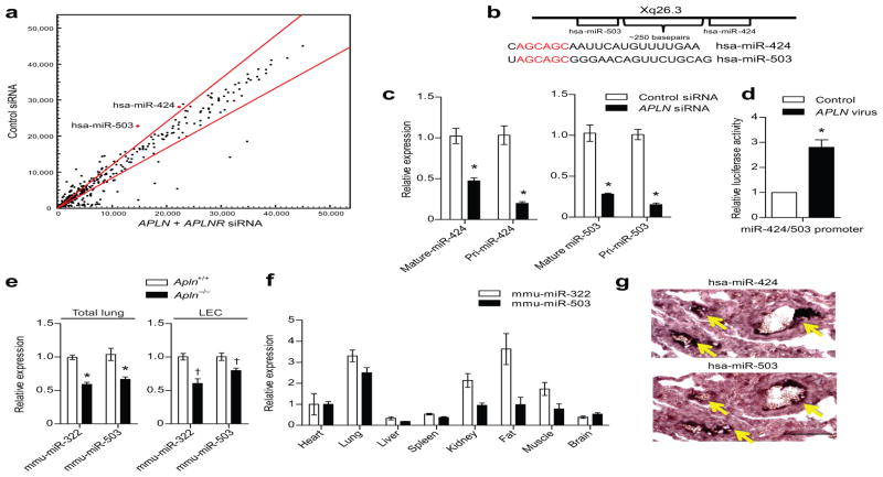
Figure 2. APLN-FGF2 reciprocal axis is mediated by APLN regulated expression of miR-424 and miR-503.
(a) MiRNA microarray analysis of PAECs subjected to APLN and APLNR knockdown. MiR-424 and miR-503 are depicted in red. Red lines demarcate a 1.2 fold change from baseline expression. (b) Chromosomal location of miR-424 and miR-503 and the sequences of the mature miRNAs. The homology in the seed sequences is depicted in red. (c) Quantitative PCR showing expression of the mature and pri-forms of miR-424 and miR-503 in response to APLN knockdown in PAECs. *P < 0.01 vs. control siRNA. (d) Relative luciferase activity of PAECs transfected with the putative miR-424 and miR-503 promoter based luciferase reporter construct in response to APLN overexpression. *P < 0.001. (e) Expression of mmu-miR-322 (mouse homolog of hsa-miR-424) and mmu-miR-503 expression in total lungs and LECs of Apln deficient mice. *P < 0.001 and †P < 0.01. (f) Quantitative PCR of mmu-miR-322 and mmu-miR-503 of mouse tissues. (g) In situ hybridization of human lungs for miR-424 and miR-503. Yellow arrows depict positive staining cells. Scale bar = 100 μm.

