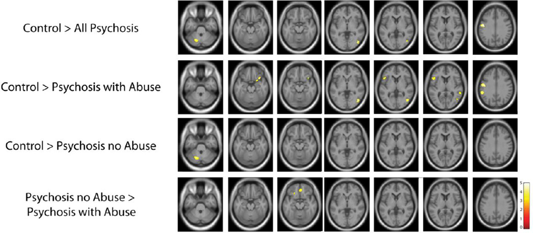
Figure 5.
Gray matter volume loss in psychotic disorder patients with and without a history of sexual abuse. Compared to healthy subjects, psychotic disorder patients demonstrated gray matter volume loss in regions consistently implicated in the disorder, including the frontal lobe, occipital lobe, and the cerebellum. Gray matter volume loss was more pronounced in psychotic disorder patients with a history of sexual abuse, with reductions across the frontal, parietal, and temporal lobes. In contrast, gray matter volume loss in psychotic disorder patients without a history of sexual abuse was significant only in the cerebellum. Comparing the two patient groups revealed bilateral frontal regions with decreased gray matter volume in patients with a history of abuse. Statistical parametric maps thresholded at a p < .001 (cluster-corrected p < .05).

