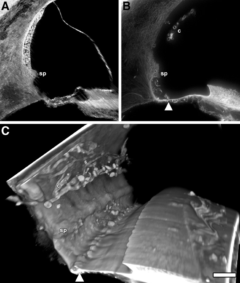
FIG. 6.
A A TSLIM scala media cross-section from a normal, formalin-fixed human temporal bone showing typical cells and tissues including the spiral prominence (sp). B SDS decellularized human temporal bone cross-section showing decellularization of scala media structures, a free-floating capillary network of the stria vascularis (c), the sp and what appears to be structures (arrowhead) on basilar membrane at a similar in location as was found in the mouse and rat. Note that there are no external sulcus cell holes in the spiral ligament similar to that which was observed in the mouse and rat. C A direct volume rendering of the scala media showing the BL of the c and sp and the basilar membrane structures (arrowhead). Bar = 100 μm.

