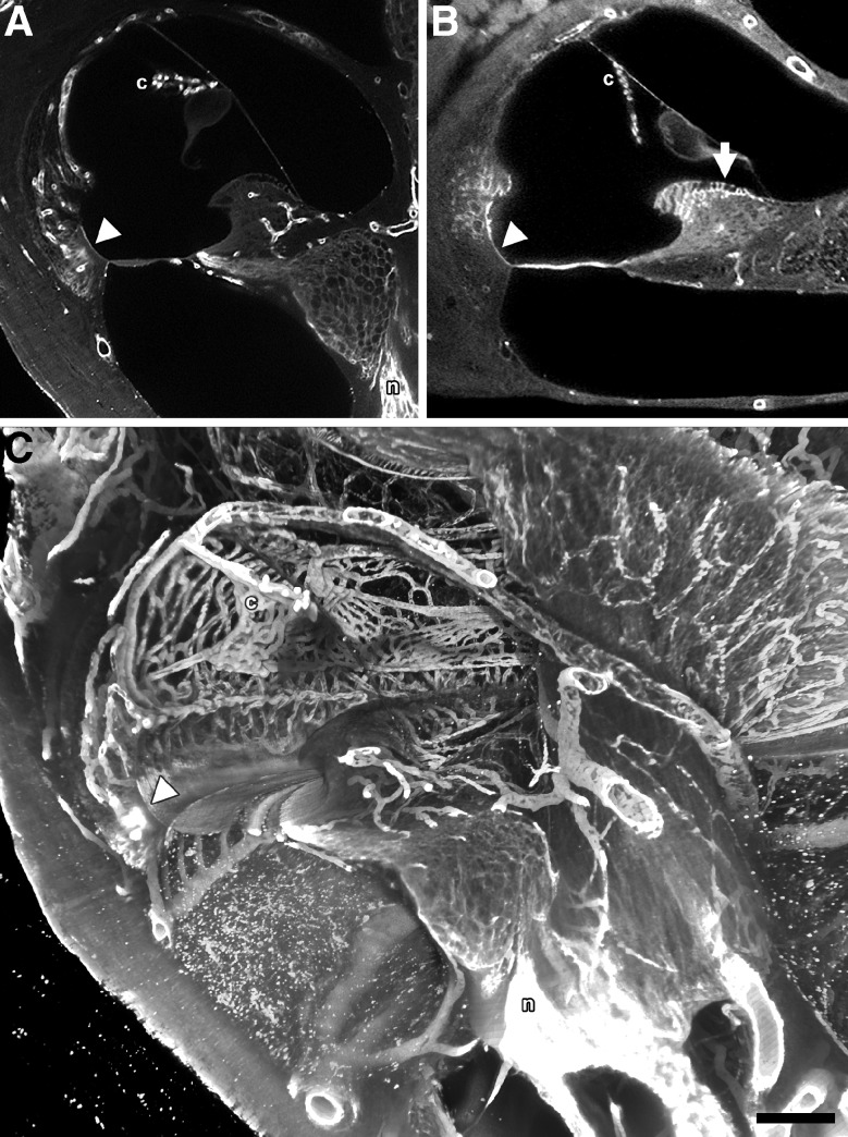
FIG. 7.
A sTSLIM optical section showing type IV collagen labeling of the BL of the capillaries of the stria vascularis (c), BL of the basilar crest (arrowhead) and the nerves (n). B sTSLIM optical section showing laminin labeling of the BL of the capillaries of the c, BL of the basilar crest (arrowhead) and BL of the interdental cells (arrow). C Direct volume rending of 50 midmodiolar sections from a collagen IV antibody-labeled, 72-h SDS-treated cochlea. Image represents a 250-μm-thick (z-dimension) volume rendering from the center of the specimen. The collagen IV antibody labeled BL in the decellularized capillaries of c, vas spirale, large vessels of the scala tympani and modiolus, spiral limbus and spiral ligament, basilar crest epithelial BL (arrowhead) as well as the antibody accessible ECM of the cochlear n. Also note that, that the scala tympani vessels appear to lie inside the scala, but they are actually encased in thin bone. Bar = 200 μm.

