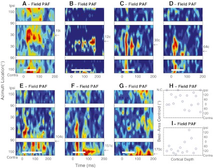
Fig. 2.
A–G PSTHs of seven units recorded in field PAF in the Idle condition. Plot conventions are the same as in Figure 1. Arrows and numbers along the right axes of the panels indicate the azimuths of the best-area centroids, expressed as ipsilateral (i) or contralateral (c) relative to the recording site. Maximum mean multiunit spike rates were 31.9, 8.6, 14.7, 9.2, 16.8, 39.3, and 24.1 spikes/s based on 36–68, 16–38, 13–34, 14–28,13–34, 14–28, and 20–32 trials at each location in A–G, respectively. H–I Best-area centroids of 18 (H) or 14 (I) units recorded along each of two probe placements in field PAF oriented approximately perpendicular to the cortical surface. NC no centroid. NC indicates that no centroid could be computed because a unit’s spike rate did not vary sufficiently across location. The ticks on the depth axis indicate intervals of 0.1 mm along the recording probes, although the specific depths in the cortex were not verified histologically.

