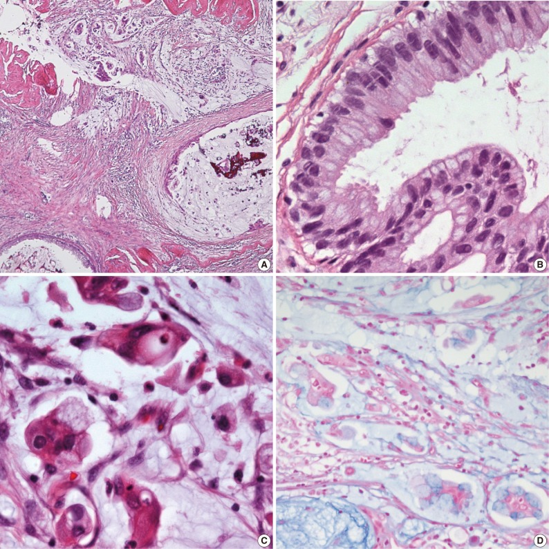
Fig. 2.
Histologic features of mucinous cystadenocarcinoma. (A) The tumor shows an abundant mucin pool in stroma with floating cell clusters. (B) The ductal carcinoma in situ is found in the form of small cysts, adjacent to the invasive carcinoma. The cysts are lined by a single layer of tall columnar mucinous cells with focal areas of micropapillary and small tufted structures, resembling those of the uterine endocervix. (C) The invasive cells are pleomorphic and contained mucin vacuoles in cytoplasm displacing atypical nuclei to the periphery. (D) Both the intracytoplasmic and extracytoplasmic mucin are stained by alcian blue.

