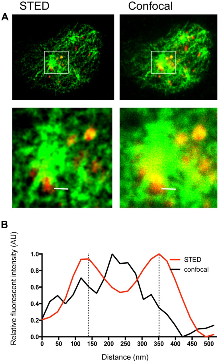
FIGURE 1.
Lytic granules on NK cell microtubules visualized by dual color STED nanoscopy and confocal microscopy. NK92 cells were activated on anti-CD18/-NKp30 coated glass as described previously (Rak et al., 2011). Cells were fixed, permeabilized and stained with tubulin biotin-streptavidin V500 (green) and perforin AlexaFluor 488 (red), then mounted with ProLong antifade. Cells were imaged on a Leica TCS confocal microscope with 100 × 1.4 NA APO objective and gated STED module. Excitation was by white light laser and fluorescence emission was detected by HyD detectors. Images were acquired and processed with LASAF software (Leica). (A) A single NK cell imaged at the plane of glass is shown in dual channel STED (left) and confocal (right). A central region is enlarged to show detail (bottom). (B) A line profile taken across the white bar shown in (A) depicts relative fluorescence of tubulin staining (arbitrary units, AU) in confocal (black) and STED (red). To generate the line profile, TIFs were exported to ImageJ (NIH) and the line profile function was used to generate raw data, which was then exported to GraphPad Prism (GraphPad Software), normalized, and graphed.

