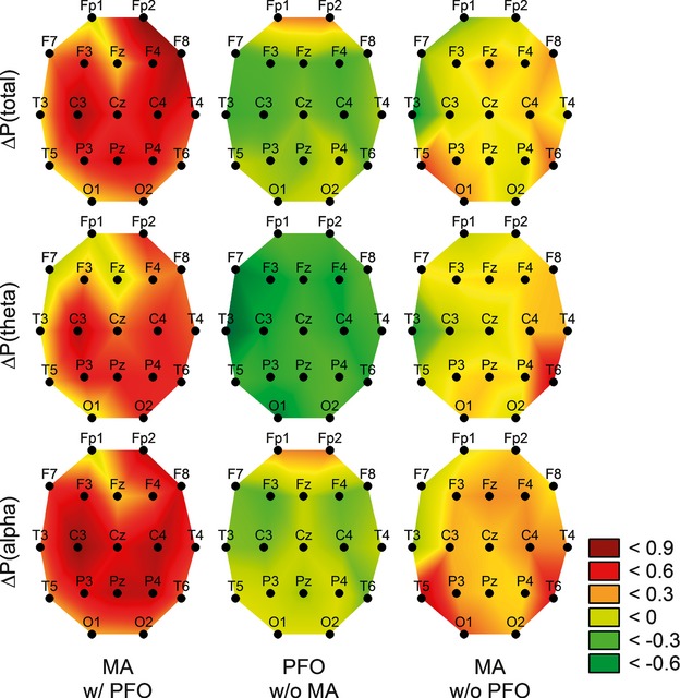
Figure 1.

Cerebral air embolism induced EEG power changes only in MA patients with PFO, not in PFO patients without migraine or MA patients without PFO. Emboli-induced spectral power changes (ΔP) were calculated for each electrode location by subtracting the baseline spectrum from the postinjection spectrum in each subject and were averaged for every group (columns). ΔP values (in decibels) are color-coded, with higher values depicting an increase. Changes in the power for total spectra (top rows) and for theta (middle rows) and alpha (bottom rows) bands are illustrated separately. MA indicates migraine with aura; PFO, patent foramen ovale.
