
Figure 12.
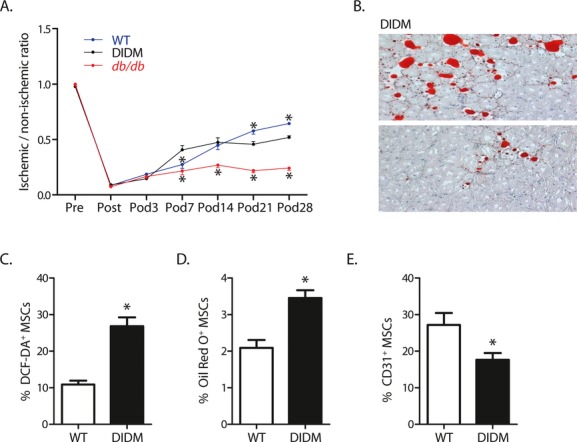
Diet-induced diabetes (DIDM) impaired recovery from hind-limb ischemia, induced adipocyte differentiation in ischemic muscle, and restricted MSC multipotency. A, Foot blood flow recovery in DIDM (mean±SEM, n=6; *P<0.05 vs DIDM; note that the WT data shown here were generated specifically for comparison with DIDM and are different from those displayed in Figure 3). B, Intramuscular adipocyte infiltration within ischemic hind-limb muscle from DIDM. C, Oxidant levels, as shown by DCF staining (n=7). D, MSCs from DIDM demonstrated increased differentiation to an adipocyte phenotype (n=8). E, MSCs from DIDM demonstrated reduced differentiation to an endothelial phenotype (n=7 to 8). (For C through E: mean±SEM; *P<0.05.) WT indicates wild-type; MSC indicates mesenchymal stem cell.