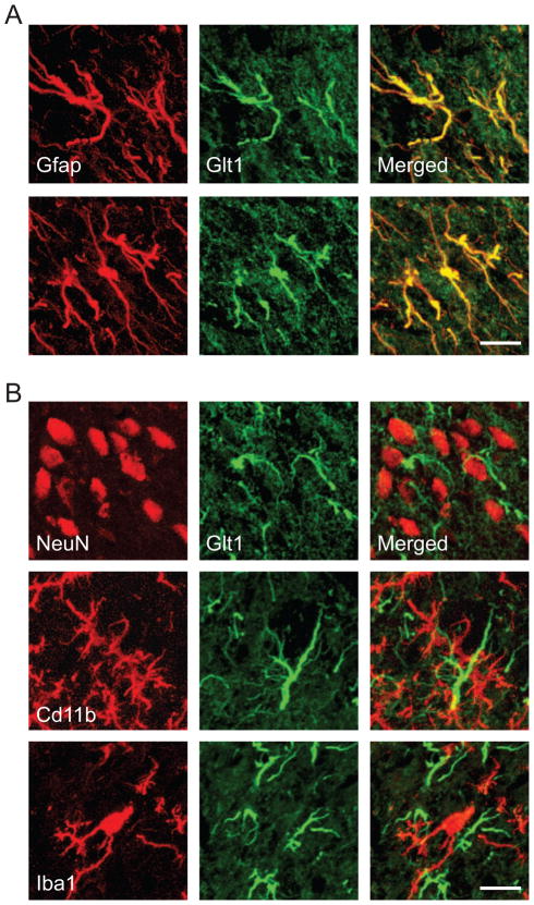
Fig. 2.
Immunohistochemical localization of Glt1 in the dorsal horn 1 week after spared nerve injury. A, Glt1 (green) was expressed by astrocytes (Gfap, red). B, Glt1 immunostaining (green) did not colocalize with neurons (NeuN, red) or microglia (CD11b, Iba1; red). The images represent z-stacks of 12–15 focal planes of 0.4 μm thickness each. Scale bars are 15 μm.

