
Fig. 1.
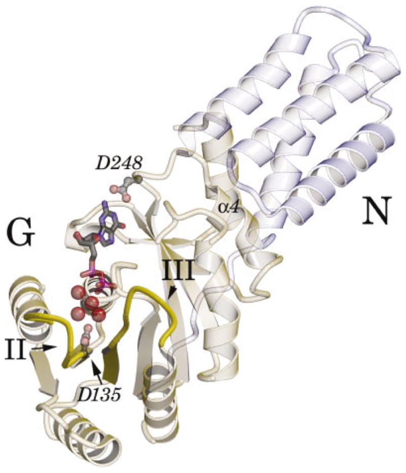
Ribbon diagram of the nucleotide bound protein. The ribbon diagram of monomer A of the Mg23GDP complex of the Ffh NG domain is oriented to view into the active site. The 3-helical N domain is in light blue, and the G domain is in light green. The motif I P-loop at the center of the G domain interacts with the phosphate groups of GDP (ball-and-stick). Motif II to the left, and motif III to the center right (indicated), interact with the bound magnesium ion through intervening water molecules. The hydrated magnesium ion is shown as a CPK representation. The carboxylate group of Asp248 hydrogen-bonds the guanine base; the carboxylate of Asp135 contributes to the second coordination sphere of the magnesium in monomer A, but is usually found in a different conformation (see text).