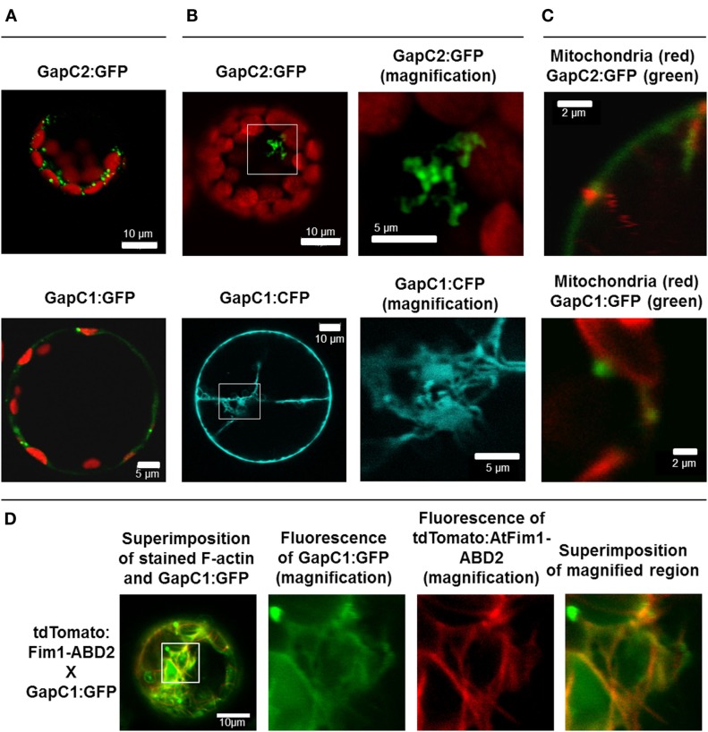
Figure 1.
Manifold localization patterns of the GAPDH isoforms. GFP- and CFP fusions of the two isoforms of GAPDH (GapC1 and GapC2) were transiently expressed in the protoplasts, isolated from leaves of 6-week-old plants of A. thaliana. (A) Superimposition of chlorophyll autofluorescence and locally accumulated GFP-fusions of the two isoforms. (B) GFP- and CFP-fluorescence from the GapC1 and GapC2 fusions, respectively. On the right hand side, magnifications are shown of relevant parts. (C) Colocalization of GFP-fused GapC1 and GapC2 with MitoTracker-stained mitochondria. (A–C). Pictures in the last lane present the magnified areas indicated in the overlays. Images in the YFP and MitoTracker channel were taken in the frame mode, both channels separately which caused a short time delay between the red and green channel, due to respective dichroic mirror settings. (D) Protoplasts were transformed with GapC1:GFP (green) and tdTomato:Fim1ABD2 (red). The fotos in the panels 2, 3, and 4 show the magnification of the boxed area in foto 1. All images were taken with the confocal Laser Scanning Microscope LSM 510 META, Zeiss.

