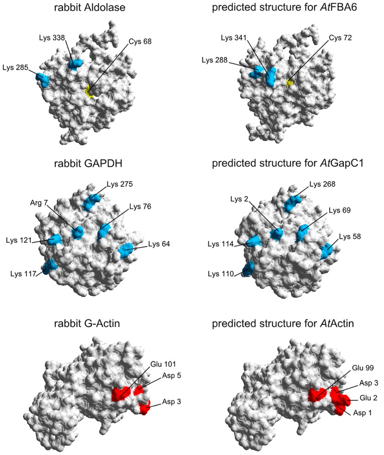
Figure 8.
Homology modeling of 3D-structures of aldolase, GAPDH, and actin. On the left hand side, the proteins from rabbit as used by Forlemu et al. (2011) are shown. The corresponding Arabidopsis proteins on the right hand side were modeled with SWISS-MODEL as described in Section Experimental Procedures. By using the Swiss-Pdb Viewer 4.0.4, the molecular surface of the generated protein models was calculated and the conserved amino acids which are necessary for the protein–protein interaction are colored: Positively charged amino acids are blue, negatively charged amino acids are red. Cysteines are shown in yellow.

