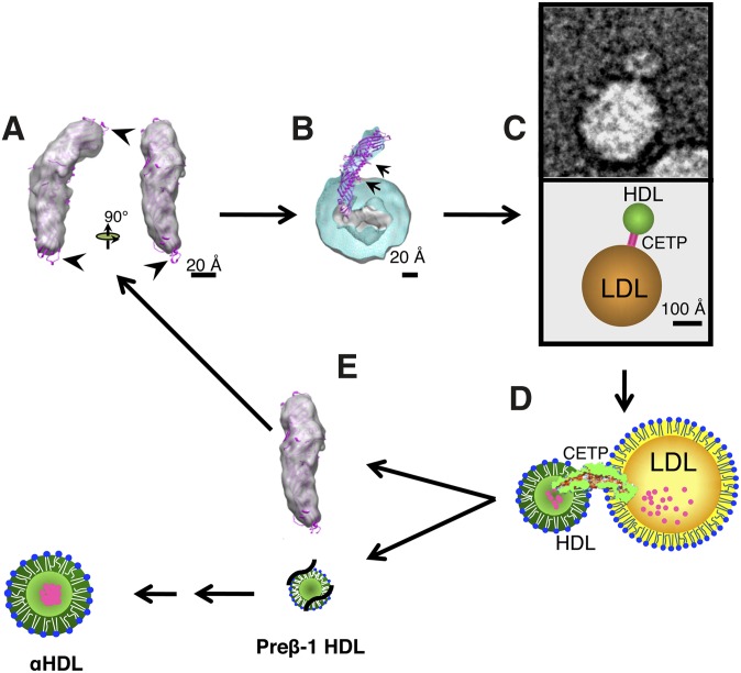
Fig. 1.
CETP's molecular and functional mechanisms (2). (A) Sensing. Three-dimensional EM molecular envelope of CETP shown in gray, and the X-ray crystal structure of CETP overlay is shown in magenta. Arrows indicate free loops extending out of the electron-dense region of CETP, with one lower loop showing the mini helix. (B) Penetration and docking. Cross-section of three-dimensional EM molecular envelope of CETP penetrating into the CE core (gray) of spherical rHDL (turquoise), with overlay of X-ray crystal structure of CETP (magenta) and arrows indicating the positions of each phospholipid pore; lower arrow also indicates the region of α-helix X. (C) Ternary complex formation. EM image (top) and diagram (bottom) of ternary complex showing CETP bridging the smaller spherical rHDL particle and the larger spherical LDL particle. (D) Lipid exchange. Illustration of CETP's interactions with lipoproteins in the ternary complex and resulting lipid transfer, in which small magenta circles represent CE moving from the smaller HDL particle to the larger LDL particle. We propose the following sequence of events: i) CETP's binding to lipoproteins may create forces resulting in the twisting of the two distal domains of CETP. ii) Twisting is the key to opening the tunnel for lipid exchange between lipoproteins. iii) Neutral lipid, e.g., CE, would migrate through the hydrophobic tunnel structure. iv) Flow and direction of lipid transfer would be established by the following dominant thermodynamic forces: differential CE concentrations, changes in hydrophobicity within CETP's central cavity favoring CE transfer from the N-terminal to C-terminal domain, and entropic energy created by the chemical potential of CE in HDL, by reason of its more ordered packing, forces CE to the less ordered (molten liquid) CE-rich (LDL) or triglyceride-rich (VLDL) lipoproteins. (E) HDL dissociation. Illustration showing the dissociation of HDL into free CETP (gray), and dissociation of relatively CE-free HDL resulting in the formation of preβ-1 HDL (black lines and small lipid structure), which is subsequently converted (arrows) into maturing α-HDL species.

