Figure 5.
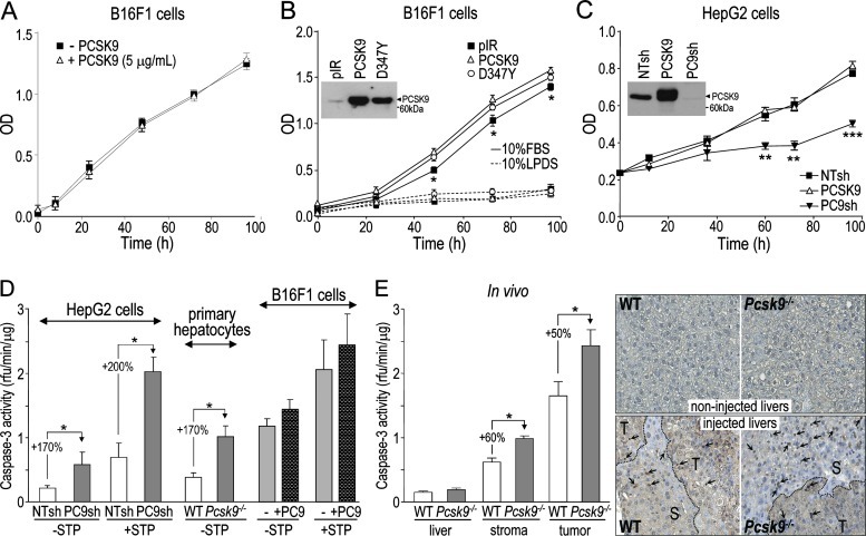
PCSK9 affects cell proliferation and apoptosis of hepatic cells but not those of B16F1 cells. (A) Proliferation of B16F1 cells incubated with or without 5 µg/ml of purified PCSK9, and grown in media containing 10% FBS, was measured using an MTS assay. (B) B16F1 cells stably expressing the empty pIRES2-EGFP vector (pIR), V5-tagged human PCSK9 (PCSK9), or its variant PCSK9D374Y (D347Y) were grown in media containing 10% FBS or 10% LPDS and their proliferation was measured. PCSK9 expression in these cell lines was assessed by Western blot analysis of serum-free media using a V5 Ab (inset). (C) HepG2 cells stably expressing NTsh, PC9sh, or human PCSK9 were grown in media containing 10% FBS and their proliferation was measured. PCSK9 expression and efficiency of PCSK9 knockdown in these cell lines were assessed by Western blot analysis of serum-free media using a V5 Ab (inset). (D) Caspase-3 activity was measured in HepG2 cells stably expressing NTsh and PC9sh, WT and Pcsk9-/- primary hepatocytes, and B16F1 cells pre-incubated with or without 5 µg/ml of purified PCSK9 for 24 hours, either in the absence or presence of 1 µM staurosporine. (E) Caspase-3 activity was assessed in non-injected liver extracts and in liver stroma and tumor extracts obtained from mice 12 days post-B16F1 cell injection. Liver sections from both non-injected and B16F1 cell-injected WT and Pcsk9-/- mice were submitted to a TUNEL assay. Arrows point at labeled apoptotic nuclei and dashed lines separate tumoral (T) and stromal (S) areas. The error bars indicate SEM of three independent experiments (A–D) or data for six mice per genotype (E). *P < .05, **P < .01, ***P < .001 (Student's t test).

