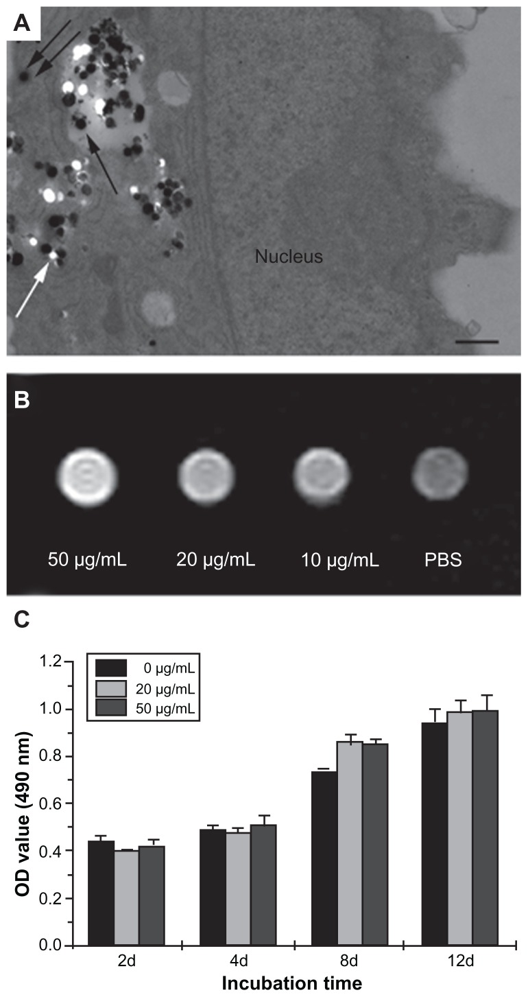
Figure 2.
(A) Transmission electron microscopy images of gadolinium-doped mesoporous silica MCM-41 (Gd2O3@MCM-41)-labeled mesenchymal stem cells (MSCs). Gd2O3@MCM-41 nanoparticles are observed as black particles in the secondary lysosome (black arrow) and the cytoplasm (double black arrows). The white spots (white arrow) were the result of the nanoparticles being separated into sections. Scale bar = 500 nm. (B) T1-weighted magnetic resonance image of MSCs incubated with Gd2O3@MCM-41 at different concentrations (50, 20, and 10 μg/mL). (C) The proliferation of Gd2O3@MCM-41-labeled MSCs at different concentrations (50 and 20 μg/mL) and non-labeled MSCs by MTT assay after 2, 4, 8, and 12 days of treatment.
Abbreviations: PBS, phosphate-buffered saline; OD, optical density; MTT, 3-(4,5- dimethylthiazol-2-yl)-2,5-diphenyltetrazolium bromide.

