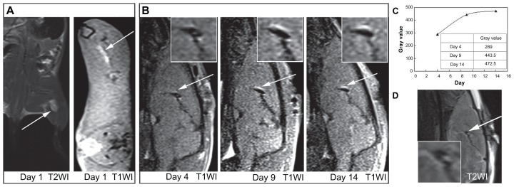
Figure 4.
(A) Magnetic resonance (MR) images of gadolinium-doped mesoporous silica MCM-41 (Gd2O3@MCM-41)-labeled mesenchymal stem cells (4 × 10⁶, 50 μg Gd2O3@ MCM-41/mL) injected into the rat thigh at 3.0 T. (B) Serial T1-weighted MR images of Gd2O3@MCM-41 labeled neural stem cells (1 × 10⁵, 50 μg Gd2O3@MCM-41/mL) injected into the rat brain on day 4, day 9, and day 14 at 3.0 T. Arrows mark the injection site. (C) The time-increased signal of (B). (D)T2-weighted MR image on day 4 showed that the signal did not come from hemorrhage.

