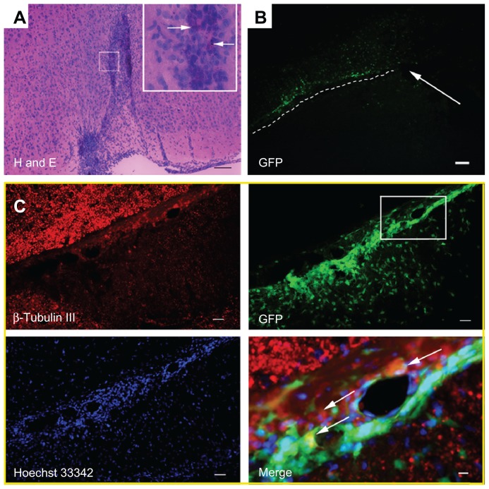
Figure 5.
(A–C) Brains of the rats were removed at 15 days posttransplantation. (A) Hematoxylin and eosin-stained sections of the rat brain. The injection site of gadolinium-doped mesoporous silica MCM-41 (Gd2O3@MCM-41)-labeled neural stem cells (NSCs) is displayed, and arrows mark the black Gd2O3@MCM-41 nanoparticles in cells. (B) Green fluorescent protein (GFP)-expressing Gd2O3@MCM-41 labeled NSCs in the brain were clearly seen. The injection area is marked with a white arrow and the migration of Gd2O3@MCM-41-labeled NSCs is shown by the white dotted line. (C) B-tubulin III (red) and Hoechst 33342 (blue) staining for GFP-expressing Gd2O3@MCM-41 labeled NSCs (green). The area in the white box in the GFP image is magnified in the Merge image. Donated NSCs (green) showed immunoreactivity for β-tubulin III (red) and Hoechst 33342 nuclear staining (blue), shown by white arrows in the Merge image. Scale bars: (A) 100 μm, (B) 80 μm, (C) 100 μm (except Merge image: 25 μm).

