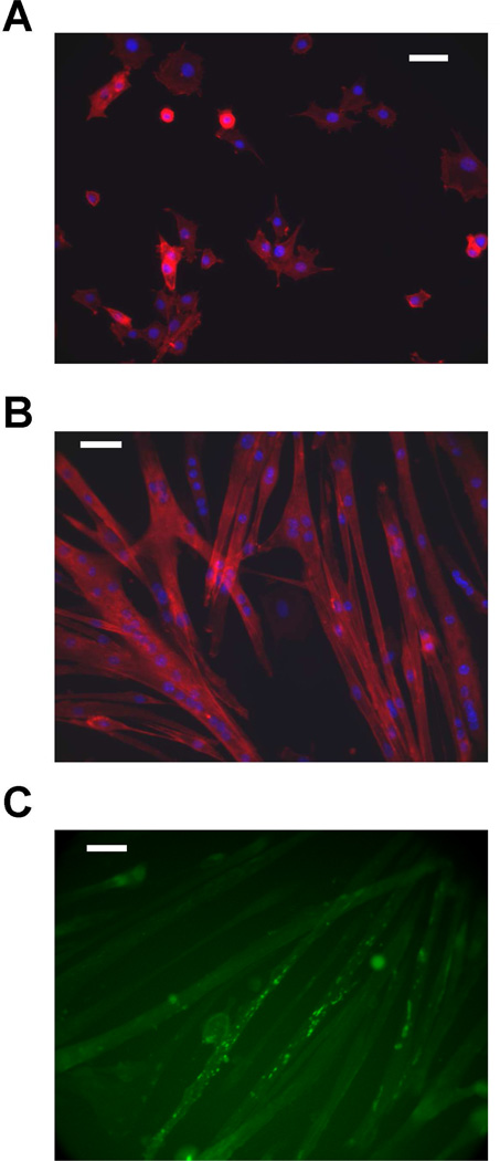
Figure 1.
Differentiation of myogenic GREG cell line in culture. (A) GREG myoblasts cultured in growth medium; nuclei stained with DAPI (blue), actin stained with rhodamine-labeled phalloidin (red) (B) formation of myotubes after 3 days in differentiation medium; nuclei stained with DAPI (blue), actin stained with rhodamine-labeled phalloidin (red). (C) nicotinic acetylcholine receptors detected by fluorescein-conjugated α-bungarotoxin. Scale bar is 200 µm.

