Figure 1.
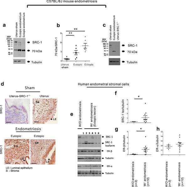
The endometriotic 70-kDa SRC-1 isoform. (a) Western blot analysis of SRC-1 expression in uterus of sham-treated mice and in the ectopic and eutopic endometrium of mice with SIE. Tubulin acts as a protein loading control. (b) The ratio of the 70-kDa fragment to intact SRC-1 in each type of endometrium (n=6/group). (c) Western blot analysis of SRC-1 expression pattern in the uterus of sham-treated mice, the eutopic endometrium of mice with SIE and the uterus of sham-treated SRC-1−/− mice. Tubulin acts as a protein loading control (d) Immunohistochemical analysis of SRC-1 expression pattern in uterus of SRC-1−/− mice, uterus of sham-treated mice and in the ectopic and eutopic endometrium of mice with SIE. (e) Western blot analysis of expression patterns of SRC-1, the 70-kDa SRC-1 isoform, ER-β and ER-α in cultured primary HESCs lines isolated from women without endometriosis (n=1) and from the ectopic lesions of women with endometriosis (n=6; lanes 1 to 6). Tubulin acts as a protein loading control. (f–h) The ratios of the SRC-1 isoform to tubulin (f), ER-β to tubulin (g) and ER-α to tubulin (h) in cultured primary HESC lines isolated from women without endometriosis (n=3) and from the ectopic and eutopic endometrium of endometriosis patients (n=10). * P<0.05, ** P<0.01 by Student's t test. Scale bars, 50 μm.

