
Figure 2.
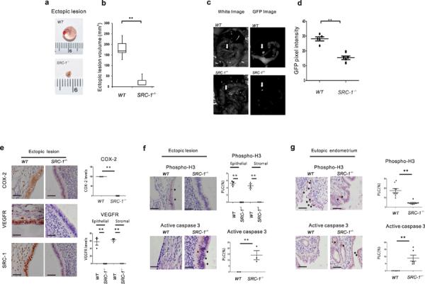
The SRC-1 gene had an essential role in the progression of endometriosis. (a) Wild-type ectopic lesions and SRC-1−/− ectopic lesions isolated from C57BL/6J recipient mice after the induction of endometriosis. (b) The volume analysis of wild-type and SRC-1−/− ectopic lesions (n=4/group). (c) The GFP images analysis of a wild-type and an SRC-1−/− ectopic lesion expressing GFP in mice with SIE. (d) The GFP pixel density of wild-type and SRC-1−/− ectopic lesions expressing GFP in mice with SIE. (e) Immunohistochemical analysis of COX-2, VEGFR and SRC-1 expression levels in wild-type ectopic lesions and SRC-1−/− ectopic lesions and quantification of COX-2 and VEGFR expression levels in each type of ectopic lesion (n=3/group). (f,g) Immunohistochemical analysis of expression levels of phospho-histone (H) 3 and activated caspase 3 in wild-type and SRC-1−/− ectopic lesions (f) or in wild-type and SRC-1−/− eutopic endometrium (g) and quantification of their expression levels in each type of ectopic lesion (n=3/group) and eutopic endometrium (n=9/group). PLC, percentage of labeled cells, **P<0.01 by Student's t test. Scale bars, 50 μm.