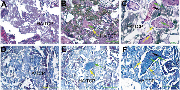
Figure 4.
Histological analysis of bone formation and trichrome staining in ceramic scaffolds seeded with iPSC-TGF-beta1-RA-cell or iPSC-RA-cells and implanted in mice. (A) Tissue section of a ceramic scaffold not seeded with cells; no bone formation is evident, only the ceramic scaffold (HA/TCP) is present. (B) Tissue section of a ceramic scaffold seeded with iPSC-RA-cells shows fewer islands of bone formation scattered in the scaffold. (C) Tissue section of a ceramic scaffold seeded with iPSC-TGF-beta1-RA-cells show many large islands of bone within the scaffold. (D) Trichrome staining in a tissue section of a ceramic scaffold implanted without cells and stained with trichrome. There is no staining for collagen, a major protein component of bone indicating absence of bone in this scaffold. (E) Ceramic scaffold implanted with iPSC-RA-cells and stained with trichrome; staining for collagen (blue) is present within the thin island of bone within the scaffold confirming H and E staining for presence of bone. (F) Ceramic scaffold seeded with iPSC-TGF-beta1-RA-cells. iPSC-TGF-beta1- RA-cells deposited more bone in scaffolds than RA alone derived cells based on histological observations of the bone size within the ceramics. Trichrome staining in the bone islands is evident confirming presence of bone matrix within the islands. Osteoblasts lining bone surfaces are indicated by yellow arrows; osteocytes are indicated by green arrows and blood vessels within the newly made bone are indicated by black arrows. Osteoblasts and osteocytes were identified by their location within bone. Cells lining the surface were presumed to be osteoblasts and cells embedded in bone were assumed to represent osteocytes. Scale bar 200 μm.

