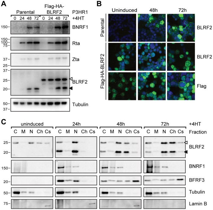
Figure 2. Characterization of BLRF2 during EBV replication.
(A) Time course of EBV protein expression using whole cell lysates from P3HR1-ZHT cells (parental) or P3HR1-ZHT cells stably expressing FLAG-HA-BLRF2 induced with 4HT for 0, 24, 48 or 72 hours. Detection of tubulin serves as loading control. Endogenous BLRF2 is indicated with a solid arrowhead and FLAG-HA-BLRF2 with an open arrowhead. (B) Immunofluorescence microscopy to determine BLRF2 localization during EBV replication in P3HR1-ZHT cells (parental, top panel) or P3HR1-ZHT cells stably expressing FLAG-HA-BLRF2 (FLAG-HA-BLRF2, bottom panels), either uninduced (left) or induced with 4-Hydroxytamoxifen (4HT) for 48 hours (middle) or 72 hours (right). Anti-BLRF2 and anti-FLAG staining are shown in green and DNA staining is shown in blue. (C) Subcellular fractionation of EBV proteins and control cell proteins from P3HR1-ZHT cells stably expressing FLAG-HA-BLRF2 induced for replication with 4HT for 0, 24, 48 or 72 hours. Equal relative amounts of the cytosol (C), membrane and organelles (M), nucleus (N), chromatin bound (Ch) and cytoskeletal (Cs) fractions were probed for the indicated proteins. Tubulin served as a control for the cytosol fraction and Lamin B for the cytoskeleton fraction. As for panel A, endogenous and FLAG-HA tagged BLRF2 are indicated with filled and open arrowheads, respectively.

