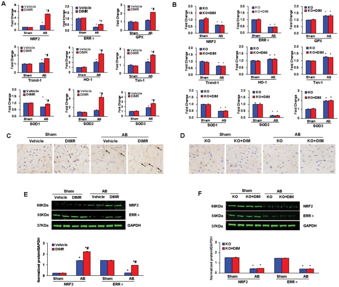
Figure 6. DIM inhibited Cardiac Oxidative Stress through AMPKα2.
(A and B) Real-time PCR analysis of the mRNA expression of ERRα, Nrf2 and its downstream genes including GPx, HO-1, Txn-1, Txnrd-1, SOD1, SOD-2 and SOD-3 in the myocardium obtained from indicated groups (n = 6). A, in WT mice and B, in AMPKα2 KO mice. (C and D) Representative immunohistochemial staining of Nrf2 protein expression in WT mice and AMPKα2 KO mice after AB. (E and F) The protein levels of the cardiac expression of Nrf2 and ERRα in the myocardium obtained from indicated groups (n = 6). E, in WT mice and F, in AMPKα2 KO mice. Top, Representative Western blots; bottom, Quantitative results. *P<0.05 compared with the corresponding sham group. # P<0.05 vs Vehicle+AB/KO+AB group.

