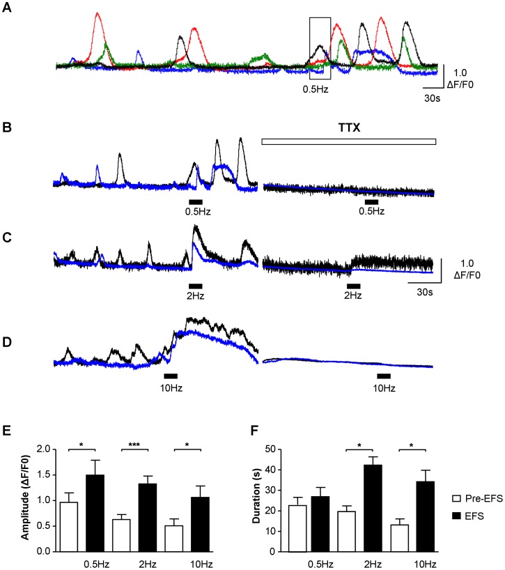
Figure 3. Functional innervation of ICC-LP. A–D.
Example of ICC-LP within a field of view responding to 0.5 Hz electrical field stimulation (EFS) where 4 cells were exhibiting non-synchronous Ca2+-oscillations pre-stimulation. EFS coordinated the activity of the 4 cells, transiently increasing their frequency of firing but not increasing the duration of the signals. Higher frequencies (2 Hz and 10 Hz) evoked simultaneous, large amplitude, long duration Ca2+-transients. EFS responses at the 3 frequencies tested were blocked by the neurotoxin, tetrodotoxin (TTX, 1 µM). (Note that traces from the 2 cells shown in panel B are from the record in panel A with corresponding colours). E, F. Summary data for amplitude and duration of pre-EFS Ca2+-oscillations and EFS-evoked Ca2+-transients, showing a significant increase by EFS (* represents P<0.05, ** represents P<0.01; *** represents P<0.001).

