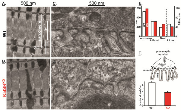
Figure 10.
KalSRKO/KO mice have abnormal skeletal muscle ultrastructure. Diaphragm muscle from WT and KalSRKO/KO male and female mice was prepared for electron microscopic analysis. Representative images showing alterations in muscle (A,B) and neuromuscular junction (C,D) architecture are shown. A sarcomere, with the A, I and Z bands marked, is shown in (A). E. KalSRKO/KO mice showed increased sarcomere (t(4)=12,p<0.001), increased I-band (t(4)=19,p<0.001) and decreased Z-band (t(4)=12,p<0.01) length. F. Junctional folds, shown schematically, were quantified per μm of junctional membrane; KalSRKO/KO mice showed a decrease in junctional fold density (t(4)=4 p<0.05).

