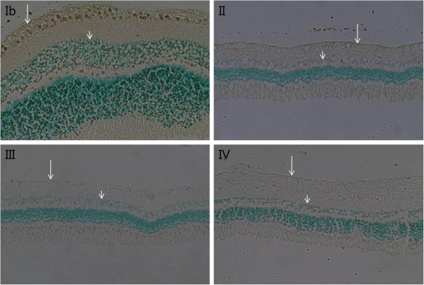
Figure 3.
Retina histological findings (TUNEL staining, x 200). An endothelin-1 dosage of 0.1 μg/day was delivered for two (II), four (III), and eight weeks (IV) and balanced salt solution was delivered for eight weeks (Ib) to the perineural region of the anterior optic nerve. Apoptotic cells (gray) were observed mainly in the ganglion cell layer (long arrow) and inner nuclear layer (short arrow) in Group Ib. However, very few apoptotic cells were observed in Groups II, III, and IV.

