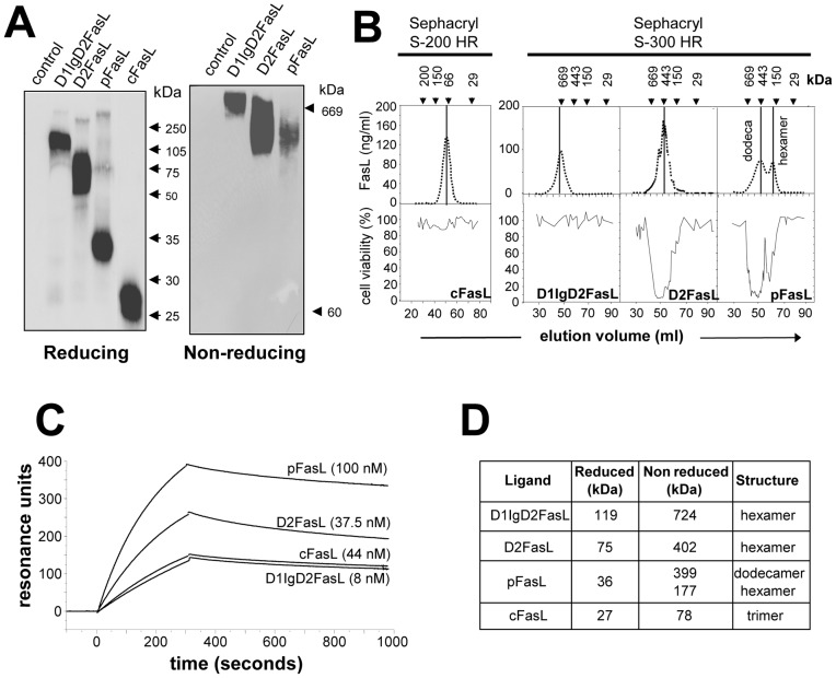
Figure 2. Biochemical characterization of the FasL/gp190 chimeras.
Panel A: Supernatants from COS cells transfected with the FasL constructs were quantified by ELISA and 10 μg of FasL protein were loaded per lane. Migrations were performed under reducing (SDS-PAGE) or non-reducing (BN-PAGE) conditions. FasL was revealed by immunoblot. Panel B: 2 μg of FasL construct were loaded on the gel filtration column. FasL was quantified by ELISA in elution fractions, and cytotoxicity was measured using the MTT assay. Panel C: Affinity measurement using Biacore®. Fas-Fc was immobilized on the chip, before the indicated soluble FasL constructs were added. A range of concentrations was tested for each analyte, but only the graph obtained with the highest concentration tested is displayed. Panel D: The apparent molecular weights and degree of oligo/polymerization of the FasL chimeras were estimated from the non denaturing gel electrophoresis and gel filtration experiments.

