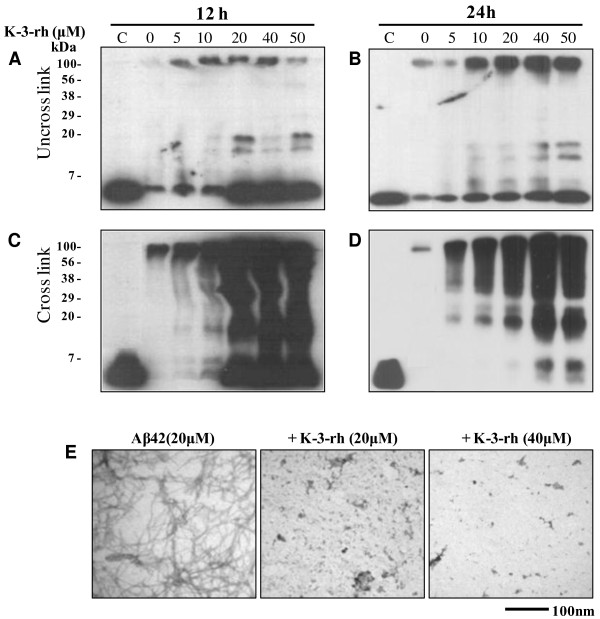
Figure 4.
Accumulation of Aβ42 aggregates in the presence of K-3-rh. Fresh Aβ42 (20 μM) was incubated in PBS at 37°C for 12 h (A and C) or 24 h (B and D), either alone or in the presence of the indicated concentration of K-3-rh. After the incubations, the peptide in the reactions was left uncrosslinked (A - B) or crosslinked (C - D) with 0.01% glutaraldehyde before being subjected to 16% SDS-PAGE and the following immunoblotting with anti-Aβ antibody 6E10. Two μl of reaction mixture was loaded for SDS-PAGE. C indicates a fresh Aβ42 (no incubation) as control. The numbers on the left indicate the relative molecular weights of protein markers. E. TEM study of the effects of K-3-rh on Aβ42 fibrillogenesis. Twenty μM fresh Aβ42 either alone (left) or in the presence of 20 μM (middle) or 40 μM (right) of K-3-rh were incubated in PBS at 37°C for 12 h. Aβ42 morphology was then visualized by TEM. Scale bar is shown at the bottom.

