Figure 5.
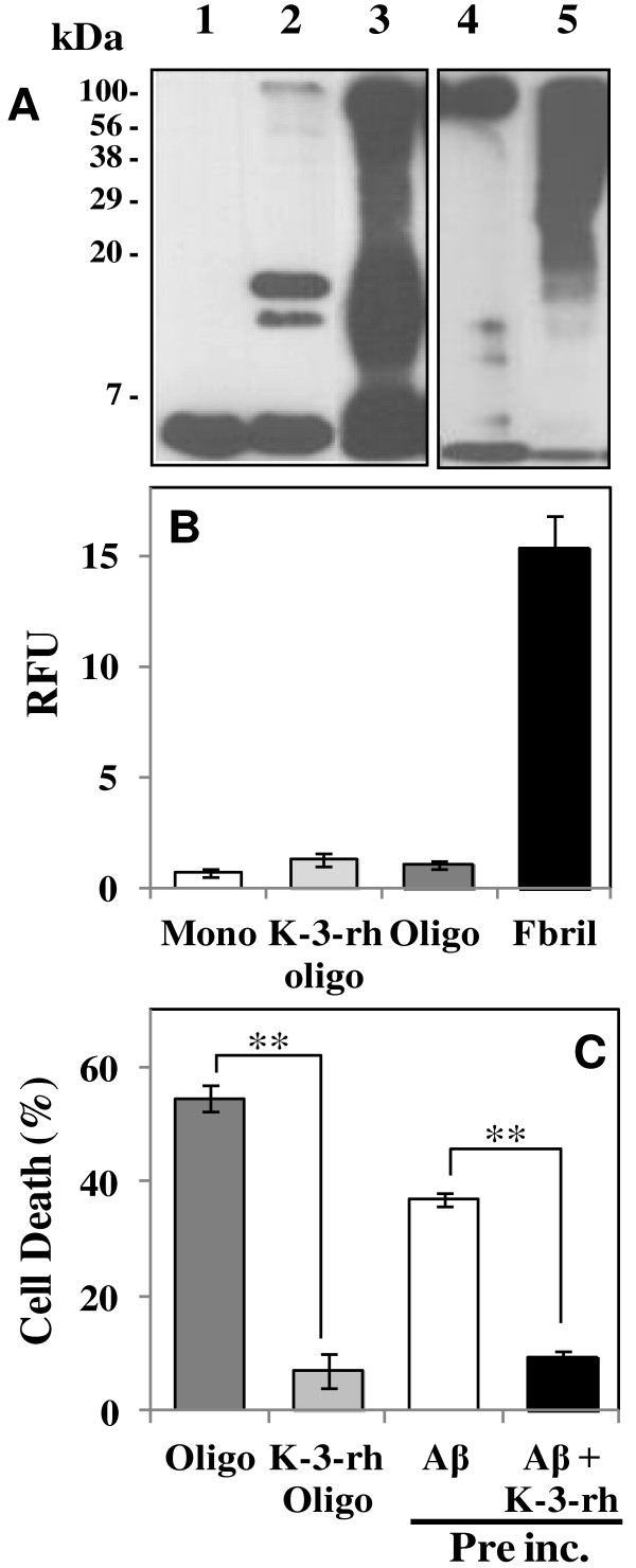
Biochemical characterization of accumulated Aβ42 species. A. Immunoblot analysis of Aβ42 oligomers probed with the 6E10 monoclonal antibody: lane 1, fresh Aβ42 as a control; lane 2, K-3-rh accumulated Aβ42 oligomers, obtained in soluble fraction by centrifuging Aβ42(20 μM) and K-3-rh(40 μM) preincubated(12 h) sample; lane 3, Aβ42 of lane 2 cross-linked before the immunoblot assay; lane 4, Aβ42 subjected to the oligomerization process and recovered in the soluble fraction after centrifugation of the mixture(Aβ42 preformed oligomers); lane 5, Aβ42 of lane 4 cross-linked before the immunoblot assay. B. Thioflavin T assay of fresh monomeric Aβ42 as control (Mono - white bar), K-3-rh accumulated Aβ42 oligomers (K-3-rh Oligo - light gray bar), Aβ42 preformed oligomers (Oligo - dark gray bar) and Aβ42 mature fibrils (Fibril - black bar). C. Decrease of the viability of SH-SY5Y cells (referred to as% of cell death) by Aβ42 preformed oligomers (Oligo - dark gray bar), K-3-rh accumulated Aβ42 oligomers (K-3-rh Oligo - light gray bar) and 20 μM Aβ42 pre-incubated in the absence (Aβ - white bar) or in the presence of 40 μM K-3-rh (Aβ + K-3-rh - black bar) for 12 h. Error bars indicate the standard deviation of triplicate independent experiments and ** indicate significant different between the groups at p < 0.01.

