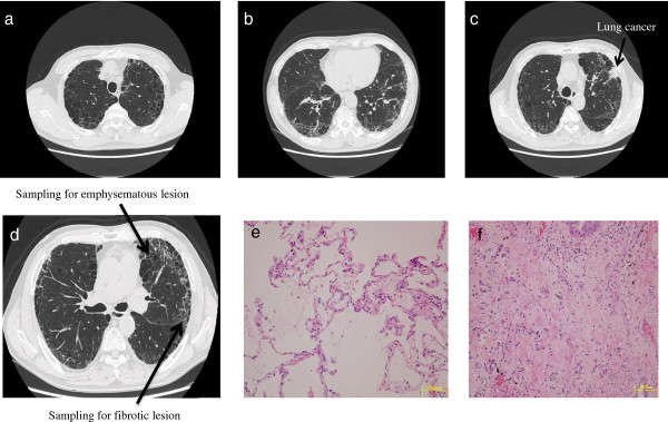
Figure 3.
Chest computed tomography and H & E staining microscopic images for case 3. Upper panel: emphysematous change in the upper zone of the lung (a), fibrotic lesion at the lung base (b), and the cancer shadow at the left superior lobe (c). Lower panel: positions (arrows) of sampling in the emphysematous lesion and the fibrotic lesion in the left superior lobe without cancerous lesions (d), and the H & E staining microscopic images of specimens of the emphysematous lesion (e) and the fibrotic lesion (f).

