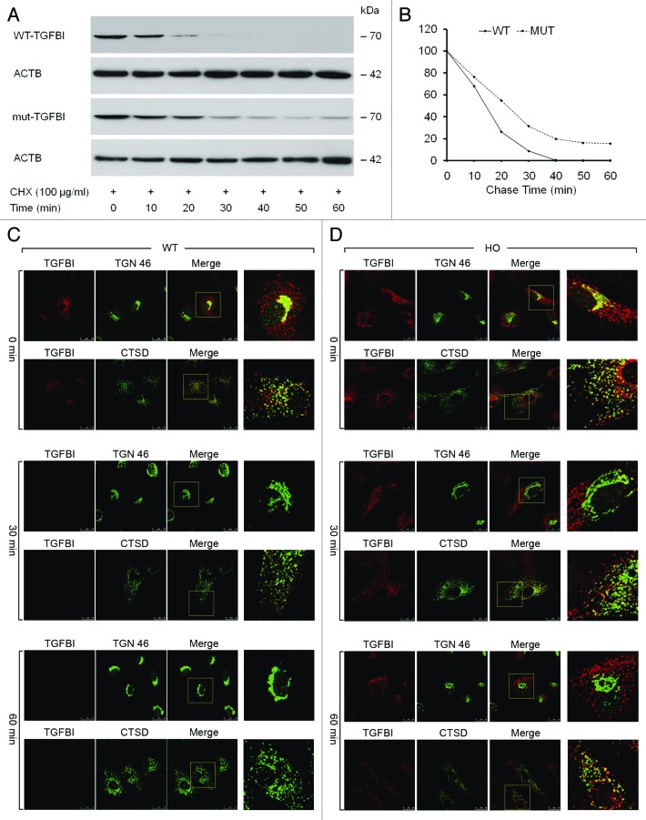
Figure 2. Kinetics of TGFBI synthesis and secretion in WT and GCD2 corneal fibroblasts. (A) Corneal fibroblasts were incubated with 100 μg/ml CHX for the indicated time periods, and the cells were then harvested and subjected to immunoblot analysis using an anti-TGFBI antibody. WT-TGFBI was not detected in corneal fibroblasts after treatment for 50 min; in contrast, mut-TGFBI remained in corneal fibroblasts for up to 60 min after treatment. (B) Most TGFBI was secreted by WT corneal fibroblasts after 40 min. However, secretion of mut-TGFBI from GCD2 homozygous corneal fibroblasts was incomplete after 60 min. (C and D) mut-TGFBI accumulates intracellularly. WT and GCD2 HO corneal fibroblasts were incubated with 100 μg/ml CHX for the indicated times and then fixed and double stained with anti-TGFBI, anti-TGN 46 and anti-CTSD. Visualization was performed with FITC-conjugated goat anti-rat IgG for TGN 46 and CTSD and with rhodamine-conjugated goat anti-rabbit IgG for TGFBI. Images were obtained with a scanning laser confocal microscope separately for TGFBI, TGN 46 and CTSD. In the combined FITC and rhodamine images, the yellow color indicates overlap between the red and green fluorescent secondary antibodies. Both WT- and mut-TGFBI colocalized with the TGN and lysosomal compartments. (C) WT-TGFBI completely disappeared after 60 min, and it could not be observed inside the cell. (D) In contrast, mut-TGFBI was colocalized with lysosomes and was maintained even after 60 min.

An official website of the United States government
Here's how you know
Official websites use .gov
A
.gov website belongs to an official
government organization in the United States.
Secure .gov websites use HTTPS
A lock (
) or https:// means you've safely
connected to the .gov website. Share sensitive
information only on official, secure websites.
