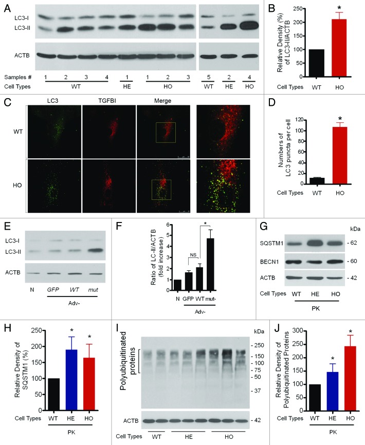
Figure 4. Assessment of levels of autophagic vacuoles in GCD2 corneal fibroblasts. (A and B) At 60–80% confluence, cells were used for LC3 western blotting analysis. Representative LC3 blots and densitometric analysis of the LC3-II/ACTB ratio using samples from wild-type (WT), heterozygous (HE) and homozygous (HO) corneal fibroblasts (three independent experiments). (C) At 60–80% confluence, cells were used for LC3 staining. Representative pictures of WT and HO corneal fibroblasts expressing endogenous LC3. WT and homozygous corneal fibroblasts grown in complete media were fixed, permeabilized and immunostained with a monoclonal antibody to endogenous TGFBI (red) and a polyclonal antibody to endogenous LC3 (green). LC3 showed vesicular staining, and the number of LC3-positive puncta was higher in GCD2 HO corneal fibroblasts compared with control cells. Insets show a 3.5-fold magnification of the indicated region. A merged image of the red and green channels is shown in the third picture in each row; yellow indicates overlapping localization. (D) Endogenous LC3 puncta per cell were quantified from WT and GCD2 corneal fibroblasts. (E) Higher LC3-II levels in corneal fibroblasts overexpressing mut-TGFBI via adenovirus-mediated gene transfer. (F) Representative LC3 blots and densitometric analysis of the LC3-II/ACTB ratio in WT, Adv-GFP, Adv-WT-TGFBI and Adv-mut-TGFBI transfected corneal fibroblasts (three independent experiments). (G) SQSTM1 and BECN1 expression levels were analyzed using anti-SQSTM1 antibody and anti-BECN1 antibody in primary cultured corneal fibroblasts (PK) from patient corneas and age-matched WT corneas. (H) Levels of total polyubiquitinated proteins were analyzed using the anti-polyubiquitinated protein polyclonal antibody in primary cultured corneal fibroblasts from patient corneas and age-matched WT corneas. (I and J) Representative western blots of polyubiquitinated proteins and SQSTM1 protein were quantified from WT and GCD2 corneal fibroblasts. Polyubiquitinated proteins, SQSTM1 and BECN1, were normalized to ACTB. Error bars: SD from three independent experiments. *p < 0.05.

An official website of the United States government
Here's how you know
Official websites use .gov
A
.gov website belongs to an official
government organization in the United States.
Secure .gov websites use HTTPS
A lock (
) or https:// means you've safely
connected to the .gov website. Share sensitive
information only on official, secure websites.
