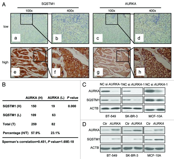
Figure 1. AURKA correlated with autophagy-associated protein SQSTM1 expression in breast cancer. (A) Both low and high expression levels of SQSTM1 immunoreactivity were mainly detected in cytoplasm and occasionally in nucleus (a, b, e and f). Both low and high expression levels of AURKA immunoreactivity were seen mainly in cytoplasm of breast cancer tissue (c, d, g and h). 100×, original magnification × 100; 400×, original magnification × 400. (B) The results were calculated on the basis of analyses performed using 341 breast cancer tissue samples. The relationship between AURKA and SQSTM1 expression was compared using Spearman’s correlation coefficient. (C) Three different human cell lines BT-549, SK-BR-3 and MCF-10A were treated with 100 nM AURKA-1 siRNA (si AURKA-1) and negative control (NC) siRNA for 24 h respectively. Cell lysates were subjected to western blot analysis with the indicated antibodies. (D) Control (Ctr) and AURKA-overexpressing cells were lysed and subjected to western blot analysis similarly.

An official website of the United States government
Here's how you know
Official websites use .gov
A
.gov website belongs to an official
government organization in the United States.
Secure .gov websites use HTTPS
A lock (
) or https:// means you've safely
connected to the .gov website. Share sensitive
information only on official, secure websites.
