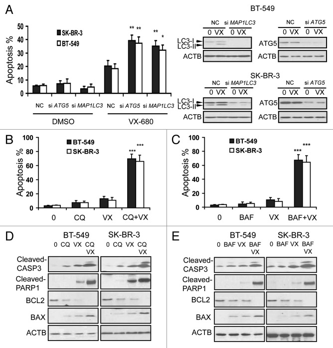
Figure 7. Inhibition of autophagy enhanced VX-680-induced apoptosis in breast cancer cells. (A) BT-549 and SK-BR-3 cells were transfected with 100nM NC siRNA, MAP1LC3 siRNA and ATG5 siRNA respectively. Twenty-four hours later, BT-549 and SK-BR-3 were treated with 10 nM and 12 nM VX-680 for 24 h respectively, cell apoptosis was measured using an Annexin V-FITC/PI staining assay. The knockdown effects on LC3 and ATG5 were confirmed by western blot analysis (right panel). (B and C) BT-549 and SK-BR-3 cells were pretreated with 100 μM chloroquine (CQ) and 200 nM bafilomycin A1 (BAF) for 1 h, and then BT-549 and SK-BR-3 cells were treated with 10 nM and 12 nM VX-680 (VX) for additional 24 h respectively, cell apoptosis was measured using an Annexin V-FITC/PI staining assay. (D and E) BT-549 and SK-BR-3 cells were pretreated with 100 μM CQ and 200 nM BAF for 1 h, and then BT-549 and SK-BR-3 cells were treated with 10 nM and 12 nM VX-680 (VX) for additional 24 h respectively, and then cell lysates were assayed by western blot analysis. Data are the means of triplicate experiments. *p < 0.05; **p < 0.01.

An official website of the United States government
Here's how you know
Official websites use .gov
A
.gov website belongs to an official
government organization in the United States.
Secure .gov websites use HTTPS
A lock (
) or https:// means you've safely
connected to the .gov website. Share sensitive
information only on official, secure websites.
