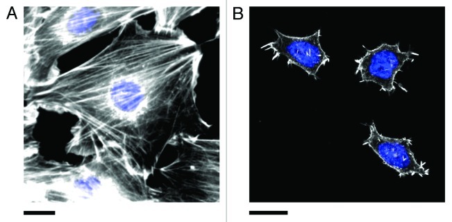
Figure 3. Actin stress fibers in (A) NIH3T3 and (B) pluripotent ES-D3 mouse embryonic stem cells (mESCs). Cells were fixed and stained for actin (phalloidin conjugated to Alexa Fluor 546, white) and the nucleus (DAPI, blue). Stress fibers are numerous and highly organized in committed NIH3T3 cells. Pluripotent mESCs (confirmed by staining for the presence of the transcription factor OCT4, not shown) are generally significantly smaller than NIH3T3 cells and display fewer stress fibers. The actin is generally diffuse and poorly organized. However, small punctate actin protrusions are observed. Experimental details in reference 11. Scale bars in both (A) and (B) are 20 μm.

An official website of the United States government
Here's how you know
Official websites use .gov
A
.gov website belongs to an official
government organization in the United States.
Secure .gov websites use HTTPS
A lock (
) or https:// means you've safely
connected to the .gov website. Share sensitive
information only on official, secure websites.
