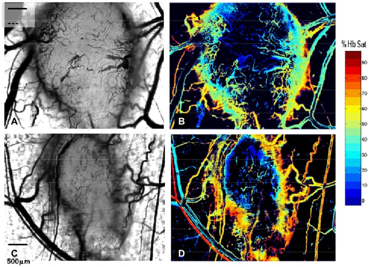
Figure 1. Eight day old 4T1 carcinoma is vascularized and hypoxic.
Intravital microscopy of two eight day old 4T1 tumors implanted in the dorsal skin window chamber viewed with light microscopy shows diffuse tumor microvascularity (panels A, C). Corresponding hyperspectral imaging of the same tumors exhibits hemoglobin saturations ≤10% over a 70% of the tumor surfaces (B,D). Magnification 5×.

