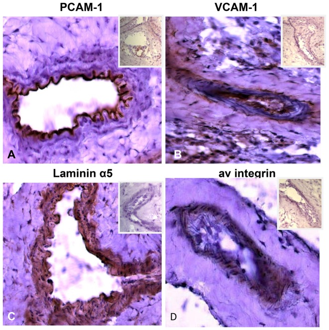
Figure 2. Expression of adhesion molecules on 4T1 tumor vascular endothelium.
Frozen sections of 4T1 tumors stained with antibodies against various adhesion molecules shows significant endothelial expression of PECAM-1 (A), ICAM-4 (B), laminin α5 (C). αv integrin (D). Secondary antibodies alone used as negative controls to stain the same tumor sections are shown in the inset of each panel. Magnification 40×.

