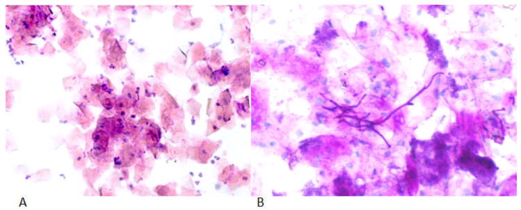
Figure 2.

PAS exfoliative cytology representative slides. (A) a healthy control benign smear; oral cytologic smear of palatal mucosa showing typical squamous cells and scattered chronic inflammatory cells. (10 X), (B) DS fungal hyphae; oral cytologic smear from palatal mucosa showing candidal hyphae. (40X).
