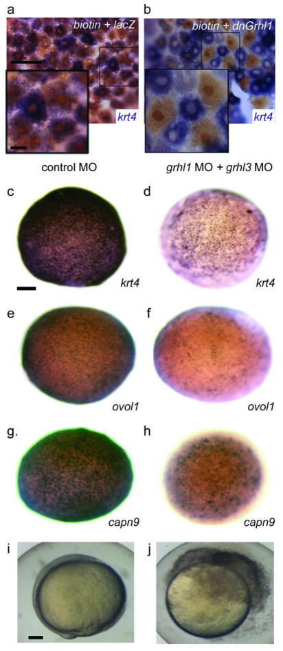
Figure 3. Knockdown of grhl family members results in loss of periderm markers.
a,b Animal-pole view of 8hpf embryos injected mosaically with a, biotinylated dextran mixed with lacZ or b, dnXgrhl1 mRNA. Embryos were fixed at shield stage and processed to reveal krt4 expression (purple) and biotin (brown). Periderm cells inheriting lacZ (brown nuclei) express krt4 variably, like periderm cells in uninjected embryos, while those inheriting dnXGrhl1 (brown nuclei) lack krt4. c–j Embryos injected with control MO (c,e,g,i), or grhl1 and grhl3 MOs (d,f,h,j), fixed at 8 hpf, and processed to reveal expression of the indicated gene, or allowed to develop until 10 hpf (i–j), at which time control MO-injected embryo have normal morphology (i), and grhl1/grhl3 MO-injected embryos have ruptured (j). Scale bars: a, 50 μm; a inset 10 μm; c, 100 μm; i, 100 μm.

