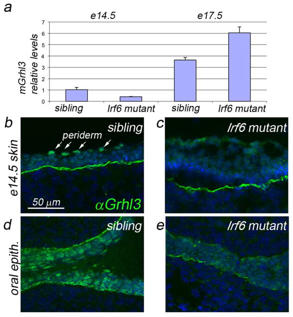
Figure 5. Expression of Grhl3 in Irf6-deficient mice.
a qRT-PCR analysis of Grhl3 levels in epidermis harvested at the indicated stage: e14.5, 2 replicates; e17.5, 3 replicates; p<0.03. b–e Anti-Grhl3 immunofluorescence on the indicated tissue at the indcates stages. In the epidermis of sibling control embryos (b), Grhl3 immunoreactivity (IR) is prominent in all epidermal and peridermal nucleii. In mutant embyros (c), Grhl3 IR is diffuse, non-nuclear, and weak to undetectable in the periderm. In the oral epithelium of control siblings (d), Grhl3 IR is strong in in all nuclei, and strongest in the oral periderm. In mutant embryos (e), Grhl3 IR is highly reduced. Scale bar: b, 50 μm.

