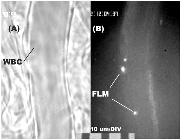
Figure 1.
Illustration of adhesion of WBCs (panel A, transmitted light) and lectin coated fluorescently labeled microspheres (FLM, panel B, fluorescence epi-illumination) to the luminal surface of post-capillary venules in mesentery of the rat. Adhesion was characterized in terms of the number of WBCs or FLMs adhering per 100 μm length of venule. The total number adhered was sampled by focusing the microscope up and down.

