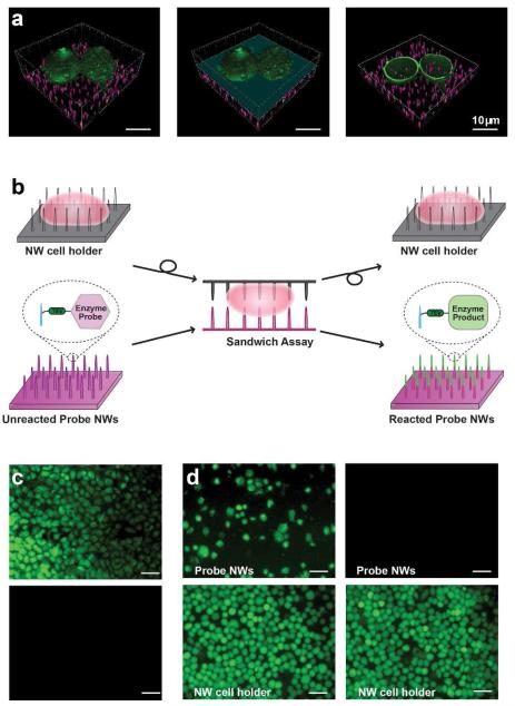
Figure 1.
(a) Confocal microscope images of membrane-labeled HeLa cells (green) on fluorescently labeled NWs (magenta). Removing the top half of the cell on NWs (left panel), as indicated by the light blue plane (middle panel), reveals NWs that reside inside the cell (right panel). Scale bar, 10 μm. (b) Schematic of the NW-cell sandwich assembly. (c) Covalent tethering of peptides to NWs prevents errant release. Fluorescein-labeled peptide molecules were either electrostatically tethered (top) or covalently attached (bottom) to probe NWs and the degree of their release into sandwiched cells was assessed after an overnight sandwich incubation. Electrostatically bound peptides were readily delivered into the cytoplasm of cells on the NW cell holder while covalently bound peptides were not transferred. Scale bar, 50 μm. (d) Validation of cell immobilization on a NW cell holder. HeLa cells, seeded in excess on the NW cell holder, were either untreated (left) or treated briefly with a TrypLE Express solution (right). After overnight sandwich incubation and disassembly, both samples were treated with CFDA for cell visualization. Weakly bound, multilayered cells were transferred from the NW cell holder to probe NWs (upper-left), whereas all cells stayed on the NW cell holder in the TrypLE Express-treated sample (note absence of cells, upper-right). Scale bar, 50 μm.

