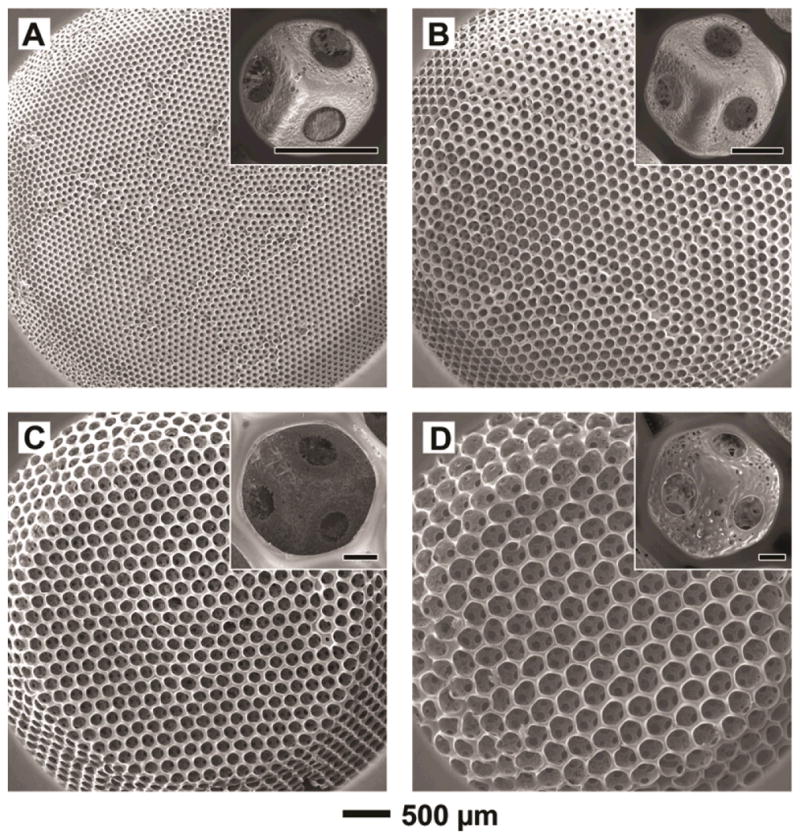
Figure 2.

(A–D) SEM images of PLGA inverse opal scaffolds with pore sizes of 79, 147, 224, and 312 μm, respectively. The scaffolds had a diameter of ~4 mm at the top surface, a diameter of ~6 mm at the bottom, and a thickness of ~1 mm. The insets show magnified views of a pore on the surface of each scaffold, revealing the uniform windows connecting to adjacent pores (scale bars: 50 μm).
