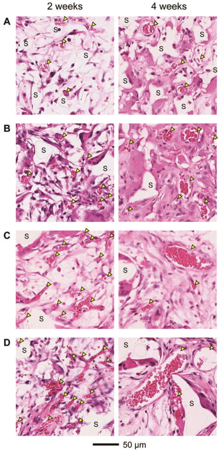
Figure 3.
Representative hematoxylin and eosin stained sections of subcutaneously implanted scaffolds with pore sizes of (A) 79, (B) 147, (C) 224, and (D) 312 μm, respectively, at 2 weeks (left column) and 4 weeks (right column) post implantation. All the images were obtained at approximately 200 μm in depth from the top surface of the scaffolds. Blood vessels are indicated by yellow arrowheads, whereas ‘S’ indicates the scaffolds.

